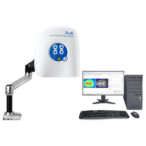
RFLSI-ZW Laser Speckle Contrast Imaging System
RFLSI-ZW Laser Speckle Contrast Imaging System - RWD
Specifications
| Resolution | Max Camera Resolution:2064×1544 pixels Best Resolution:3.9 μm/pixel |
| Image | Flux/Gray/Intensity/Color/Overlay |
| Measurement Laser | 785 nm, Class 1 |
| Indicating Laser | 650 nm×2, Class 1 |
| Focus | Auto/Manual (fine focus) |
| Trigger | 2×BNC |
| Image Size | 0.57× 0.75-22.5×30 cm2 |
| Max Frame Rate | 100 FPS (full field) |
| Zoom | 10× |
| Working Distance | 10-40 cm, continuous |
| System Calibration | Calibration Box |
| MEASUREMENT ALGORITHMS | Temporal and Spatial processing |
| PC CONNECTIONS | 1 x USB 3.0 port |
| Software | Acquisition Software and Analysis Software |
| POWER SUPPLY | Universal Voltage, 100V-230V. Note acquisition rate is unaffected by frequency of local electrical supply. |
| STAND OPTIONS | Scan head has standard VESA mount for desktop stand, Microstand and Clinical Mobile stand. |
Applications:
-
Cerebral blood perfusion monitoring
-
MCAO model assessment
-
Cortical spreading depression observation
-
Hind-limb ischemia research
-
Skin burn/skin flap transplantation
-
Organ microcirculation observation
-
Skin allergies
-
Septic Shock
-
Chicken Chorioallantoic Membrane Assay
-
Diabetic Foot
Highlights Of RFLSI-ZW
-
Image any exposed tissue (skin or surgically exposed tissues) and species.
-
Non-contact, non-contrast agent depending measurement.
- The built-in CMOS global shutter camera can achieve faster data acquisition and processing speed.
-
Best optical resolution of 3.9 μm/pixel, providing more detailed tissue structures.
-
Max frame rate (full field) up to 100 fps, acquiring real-time changes in larger areas.
-
Motorised 10x optical zoom and auto focus. Image size ranges from 0.57×0.75 to 22.5×30 cm2 in all-in-one imager, covering multiple research applications.
-
Fast auto and fine manual focus, improving focus efficiency and accuracy on various tissues.
-
Optimal lens assembly, filtering the ambient and reflecting light.
-
Class 1 of measurement and indicating lasers, safe to use without eye protection System
-
Laser stability hardware for the ultimate in reliable and consistent measurement over minutes, hours and days.
-
Calibration with calibration box. Self-calibration is possible at any time to keep the equipment in optimal working condition.
-
Trigger In/Out BNC connections for communication with external devices.
-
Unlimited installation of analysis software in PC.